Ultrasound ablation,Noninvasive medical representative,Creating happy family
HIFU & Focused Ultrasound
Ultrasound is a form of vibrational wave. It can transmit harmlessly through living tissues, and this makes it possible to use an extracorporeal source of ultrasound for therapeutic purposes. If ultrasound beams are focused and sufficient ultrasonic energy is concentrated within volume while they propagate through tissues, the temperature in the focal region may be raised to levels at which the tumors are cooked, resulting in tissue ablation. This process occurs without any damage to surrounding or overlying tissues, and the tissue ablation technique that employs such beams is known interchangeably as high intensity focused ultrasound (HIFU).
HIFU is a different therapy from conventional hyperthermia, which has been used as an adjuvant to radiotherapy and chemotherapy for cancer treatment since the 1980s. The purpose of hyperthermia to raise the temperature of the tumor from 37℃ to 42-45℃, and to maintain uniform temperature distributions in a narrow therapeutic range for 60 minutes. However, the temperature distributions induced in vivo are usually non-uniform because of tissue cooling by blood flow, and it is extremely difficult to avoid local cold spots that do not reach the necessary therapeutic temperature level. With HIFU, the temperature within the focal zone is rapidly raised to temperatures between 56℃ to 90℃ and is held for one second. The rapid deposition of HIFU thermal energy causes a peak temperature rise that is unaffected by blood flow cooling. Therefore, it can avoid the problem of non-uniform temperature distribution in hyperthermia, and there is no need for inserting thermocouple probes into the targeted tumor to describe temperature distribution during HIFU procedures.
Ultrasound Ablation
Mechanism of Ultrasound Ablation
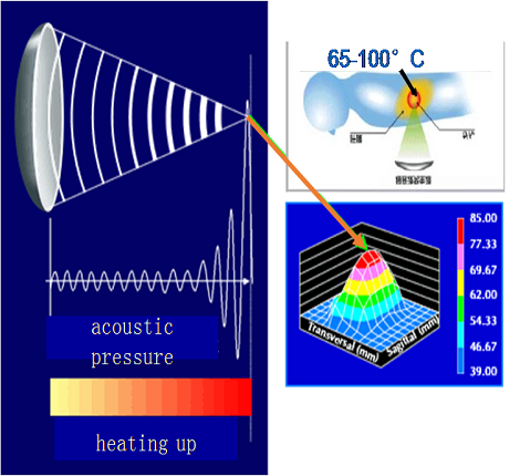
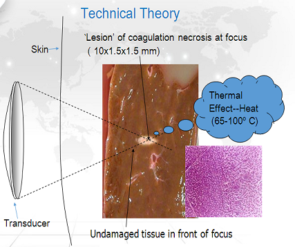
Several mechanisms are directly involved in the tissue damage induced by Ultrasound Ablation(UA). The first is a thermal effect from the conversion of mechanical energy into heat in the targeted tissue, the thermal effect depends on the temperature achieved during Ultrasound Ablation(UA). If the temperature rise is above a threshold of 56℃ and the exposure time is one second, irreversible cell death will be induced through coagulation necrosis. In fact, the temperature at a focal volume may rise rapidly above 80℃ during Ultrasound Ablation(UA).
The second is acoustic cavitation, the presence of small gaseous nuclei existing in subcellular organcelles and fluid in tissue are the source of cavitation, which can expand and contract under influence of the acoustic pressure is more than several thousand Pascals, and the temperatures reach several thousand degrees Celsius, resulting in tissue damage.
Damage to tumor vasculature, caused by Ultrasound Ablation(UA), may account for the third mechanism of the targeted tissue necrosis. It can directly cease the blood supply to the tumor through the destruction of the tumor-nourishing vessels, thus causing deprivation of nutrition and oxygen for the tumor cells, indirectly resulting in coagulation necrosis.
In fact, it seems impossible to distinguish the thermal effects from either acoustic cavitation or vessel damage in Ultrasound Ablation(UA), and they can occur simultaneously within the targeted tissue. Therefore, the coagulation necrosis induced by Ultrasound Ablation(UA) can be considered as the result of biological effects from a combination of heat, cavitation and vascular destruction on tissue.
• Risk of infection
• Post operative pain
• Hospital stay
• Recovery time
• Disfiguring
• Loss of earnings
• One time Treatment
• Real-time US(MRI) guided
• Procedure
• 3D-conformal treatment
• Anti-tumor immunity activated
• Minor complications
• Day Care basis
• Low overall cost
• Enhancement of quality of life
• Liver
• Kidney
• Breast
• Pancreas
• Bone
• Soft Tissue
• Metastatic cancers
• Uterine Fibroids
• Uterine adenomyosis
• Breast Fibroadenoma
© 2020 Chongqing Haifu Medical Technology Co., Ltd. All rights